Ordering
Email: [email protected]
Ordering: +45 29 13 42 24
Main: +45 31 10 91 91
CVR/VAT no. 31348854
Technical Support
Contact technical support
Tracking T cells in the tissue where they exert their function can advance our understanding of T-cell immunity. MHC I Dextramer® for in-situ staining reagents can detect spatial distribution, localization, and abundance of antigen-specific T cells in different tissue sites in health and disease.
Antigen-specific T cells in tissue sections can be visualized by direct or indirect immunohistochemistry (IHC) methods. Direct visualization uses the conjugated fluorophores on the MHC I Dextramer® reagent, while indirect visualization uses antibody staining directed against the fluorophore on the MHC I Dextramer® for signal amplification. MHC I Dextramer® for in-situ staining reagents provides T-cell staining in fresh samples, paraffin-embedded (FFPE), and frozen sections.
Solid tissue like tumors and lymphoid organs have different structures and spatial distributions of infiltrating T cells, often challenging to identify.
Immudex offers the Dextramer® In-Situ Staining Kit to optimize T-cell staining in solid tissue sections by IHC and comprises three Dextramer® reagents where:

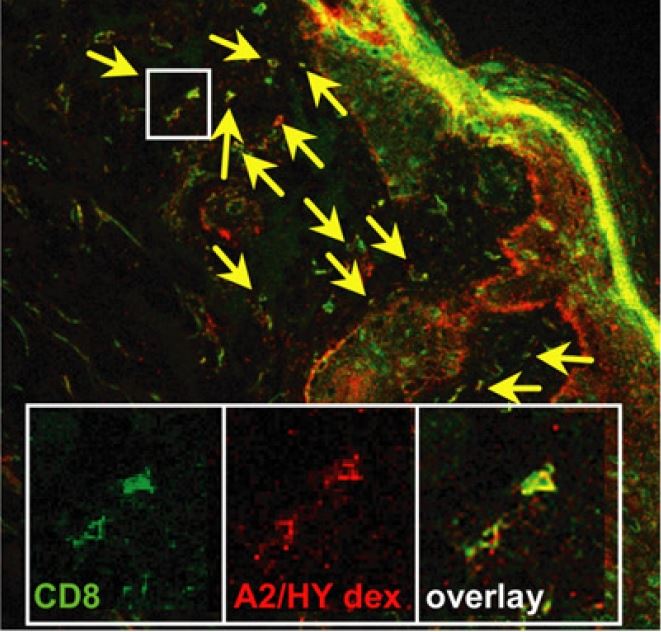
Cryosections stained with CD8 antibody (green) and HLA-A2/HY Dextramer® (red). Yellow arrows indicate colothecalization of HLA-A2/HY Dextramer® and CD8 antibodies on the same cell.
Figure adapted from Kim YH, et al., 2012
By utilizing the Dextramer® In-Situ Staining Kit, you can identify the reagent optimal for your tissue type and experimental setup. Optimization may also be required for other steps of the IHC procedure, e.g., tissue preparation and antibodies.
Immudex manufactures Dextramer® for in-situ staining reagents made with MHC class I alleles, as listed in the table below. Customized Dextramer® for in-situ staining reagents are easily made with MHC alleles from our catalog and peptides forming stable complexes with the MHC molecule.
Send an e-mail to [email protected] specifying:
| Content | Amount | Fluorophore | Price (EUR) | Price (USD) |
| Dextramer® In-Situ Staining | 0,2 mL | FITC | 3.430 | 3,910 |
| Dextramer® In-Situ Staining | 1 mL | FITC | 8.620 | 9,845 |
Find your Dextramer® reagent in the table below, or send an e-mail to [email protected] specifying:
Immudex has updated the catalog numbers. To learn more about it, please consult the document here
Ordering
Email: [email protected]
Ordering: +45 29 13 42 24
Main: +45 31 10 91 91
CVR/VAT no. 31348854
Technical Support
Contact technical support
Our webshop does not include all products. Contact us or email [email protected] and we will search our larger internal database of products for you. We also provide custom alleles and specificities.
BV421 is equivalent to Brilliant Violet™ 421, which is a trademark or registered trademark of Becton, Dickinson and Company or its affiliates, and is used under license. Powered by BD Innovation.