Get the case study as a PDF.
Dextramer® In-Situ Staining of Antigen-Specific, Autoreactive CD4+ T Cells
Background
Unlike MHC class I multimers, using MHC class II multimers for CD4+ T cells detection can be challenging, especially in the case of low-affinity cells. For the first time, this study from Massilamany et al. reports the utility of MHC II Dextramer® reagents for successful detection and quantitative analyses of antigen-specific, autoreactive CD4+ T cells in murine fresh tissue sections.

Study Description
Goal: to detect and quantify antigen-specific, autoreactive CD4+ T cells in-situ, using experimental autoimmune encephalomyelitis (EAE) and experimental autoimmune myocarditis (EAM) mouse models.
Primary T-cell cultures were derived either from SJL mice immunized with myelin proteolipid protein (PLP) 139-151 (EAE model) or A/J mice immunized with cardiac myosin heavy chain-α (Myhc) 334-352 (EAM model) and stained with the corresponding MHC II Dextramer® reagents. Autoreactive CD4+ T cells were further detected and enumerated in-situ from the the brain and heart sections by IAs/PLP 139-151 and IAk/Myhc 334-352 Dextramer® staining and laser scanning confocal microscopy (LSCM).
Results
- IAs /PLP 139-151 Dextramer® in-situ staining directly detected PLP 139-151 CD4+ T cells in the brain sections (Fig.1.)
- PLP-specific Dextramer® in-situ staining was better at room temperature than 4° or 37°C concerning staining intensity and/or specificity (data not shown)
- Detection of PLP 139-151 Dextramer®-positive CD4+ T cells varied between mice (0.8% to 3.3%) and between tissue sections in each mice (0.8% to 5.5%), as expected (data not shown).
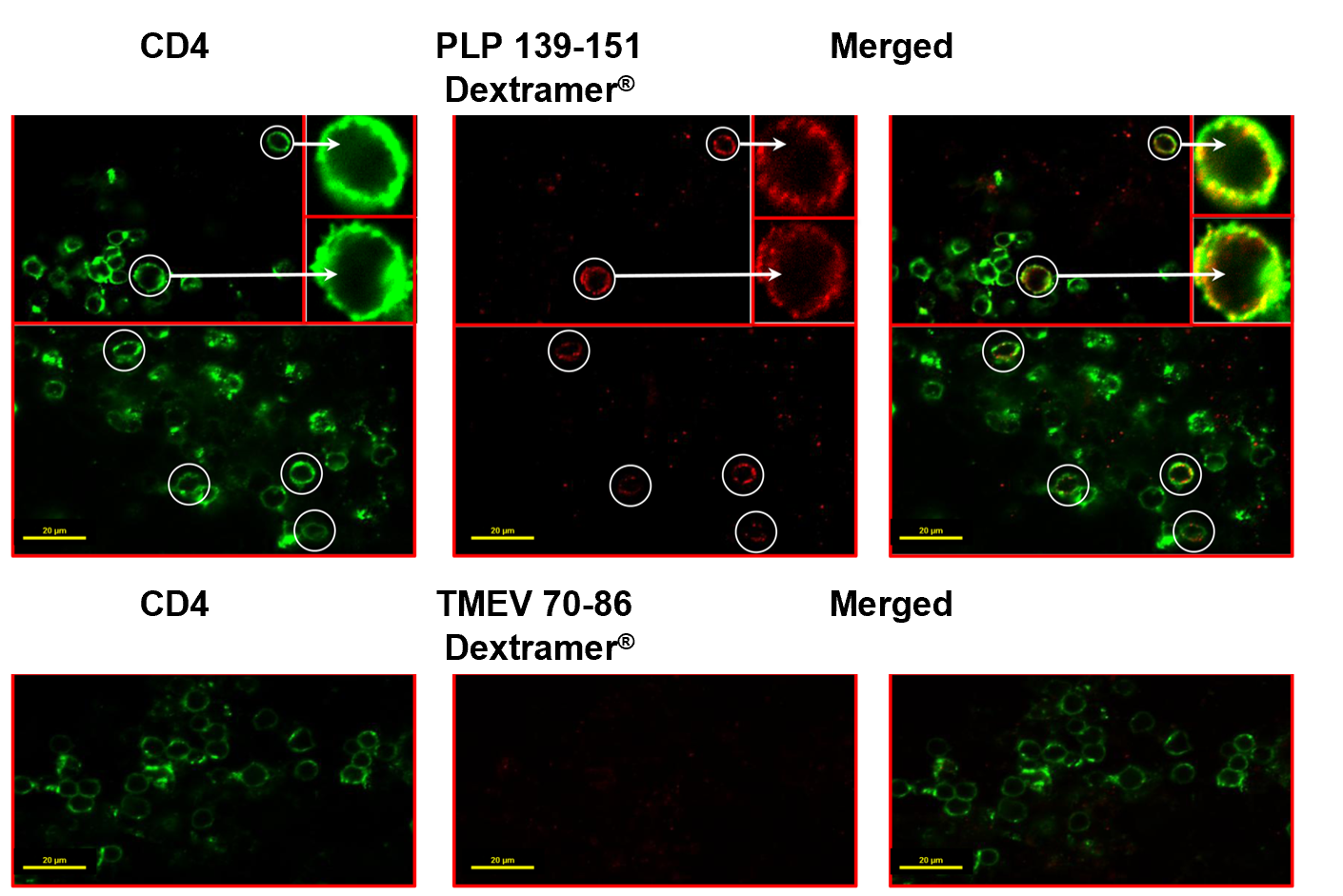
Fig.1. Detection of PLP-specific T cells by in-situ staining with IAs /PLP 139-151 Dextramer®. Cerebral sections co-stained with PLP 139–151 Dextramer® (red) and anti-CD4 (green) (top panels), or TMEV 70–86 Dextramer® (control) and anti-CD4 (green) (bottom panels), merged (yellow) (circles, dext+ CD4+ T cells; insets represent enlarged views of dext+ CD4+ T cells). Original magnification 1000×; bar = 20 µm.
Conclusions
- “MHC II Dextramer® for in-situ staining reagents can be successfully used to detect antigen-specific CD4+ T cells in fresh tissue sections with a high degree of specificity by direct staining without the need to amplify the signals with fluorophore antibodies”
- MHC II Dextramer® for in-situ staining reagents allow reliable quantification of antigen-specific, autoreactive CD4+ T cells in tissue
- “In-situ staining with MHC II Dextramer® is a one-step reaction that can be finished in less than one day.”