Get the case study as a PDF.
Validation of MAIT Cells with TCRs Engineered to Recognize HBV infected cells using MHC Dextramer®
Healy et al. Human MAIT cells endowed with HBV specificity are cytotoxic and migrate towards HBV-HCC while retaining antimicrobial functions. JHEP Rep 2021 (DOI: 10.1016/j.jhepr.2021.100318)
Background
Chronic HBV infection is a leading cause of liver cancer. T-cell receptor (TCR)-engineered T cells can be modified to recognize virus-infected and/or cancer cells and may help prevent disease development by eliminating the infection. Here, Healy et al. evaluated whether mucosal-associated invariant T cells (MAIT), a large population of unconventional T cells, could recognize and kill HBV-infected hepatocytes when engineered with an HBV-specific TCR. MAIT TCR expression was confirmed and monitored using MHC I Dextramer® reagents.
Study Description
PBMCs underwent MAIT cell expansion and were cultured under the presence of AIM-V, AB serum, and activated under the presence of anti-CD3 and IL-2 to generate ConT cells. ConT and MAIT cells were transfected with HBVs183-91 TCR mRNA by electroporation. TCR expression was quantified by staining with an HLA-A2 MHC Dextramer® loaded with FLLTRILTI peptide at 4°C for 20 min before surface antibody staining. Cells were analyzed by flow cytometry.
Results
Surface expression of the TCR on the ConT and MAIT cells was successfully assessed by flow cytometry using an HLA-A2 HBVs183-91 Dextramer® to confirm TCR expression (Fig. 1A). When sampling at 12-hour intervals, the kinetics of the TCR expression was comparable for MAIT and ConT cells, peaking 24 hours after electroporation. However, analysis of the AUC of HBV TCR expression in MAIT cells indicated higher and slightly prolonged TCR expression compared with ConT cells (Fig. 1B). Gating on HBV TCR+ cells did not reveal any significant differences between ConT and MAIT cells on a CD8+, CD4+, double-negative, or double-positive level (Fig. 1C).
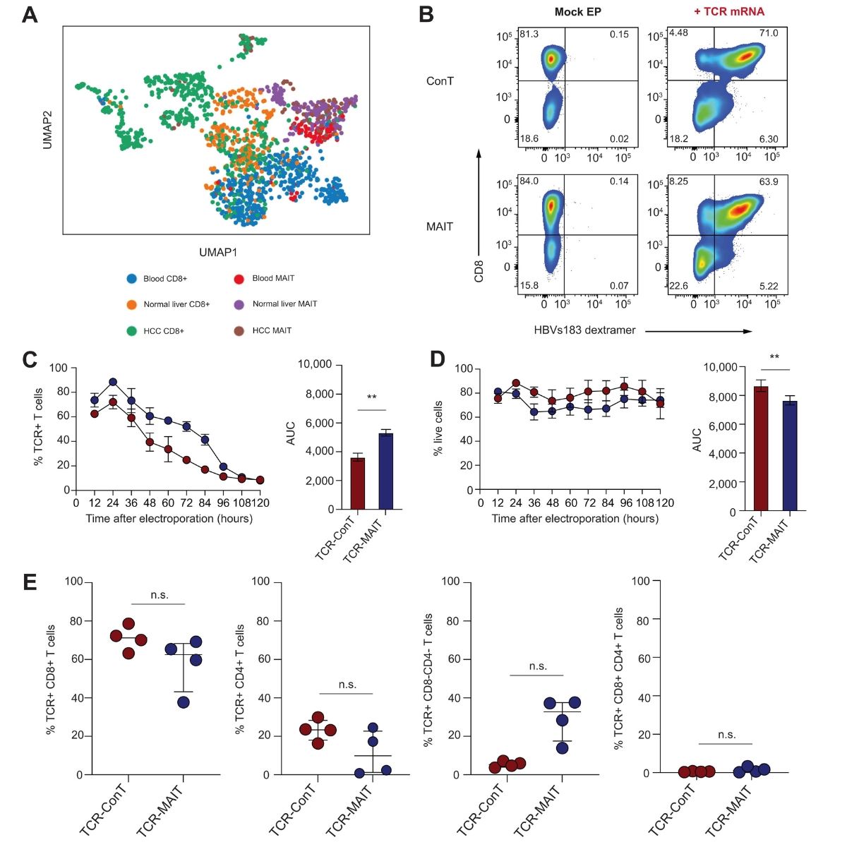
Fig.1: Production and flow cytometric analysis of HBV-specific TCR-ConT and TCR-MAIT cells. (A) Representative flow cytometry plots from a single donor of HLA-A2-HBVs183-91 Dextramer® staining in HBVs183-91 TCR mRNA electroporated ConT and MAIT cells 18 hours post electroporation. Mock electroporated T cells served as negative controls. (C) The percentage of CD8+, CD4+, CD8−CD4−, and CD8+CD4+ cells within the HBV TCR+ ConT and MAIT cell populations (n = 4). Wilcoxon matched-pairs signed-rank test was used to detect significance. ∗p <0.05, ∗∗p <0.01, ∗∗∗p <0.001.
Conclusions
- MHC Dextramer® reagents successfully detected TCR transfected MAIT and ConT cells prepared to treat HBV infection and prevent liver cancer development
- Together, these data demonstrate that blood-derived MAIT cells represent a readily accessible cell source. The MAIT cells can further be engineered with an HBV-specific TCR with high but transient efficiency for potential treatment of chronic HBV infections to prevent liver cancer.