Get the case study as a PDF.
Superior Detection of Self-Reactive, Antigen-Specific T-Cells Using Klickmer® Compared to Tetramers
Background
MHC-peptide multimers have become the “gold standard” for the detection and isolation of antigen-specific T cells. Detection sensitivity is a general issue but particularly pronounced for anticancer and autoimmune T cells. Self-reactive T-cell populations are often enriched for low-affinity T-cell receptors (TCR) due to the removal of high-affinity cells by immune tolerance mechanisms.
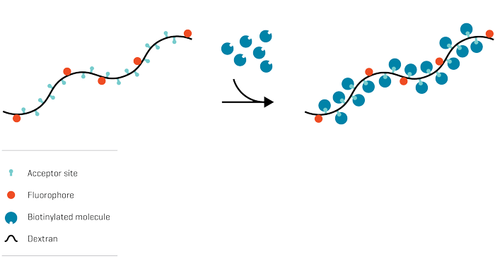
Here, Dolton et al. investigate how to optimize multimer staining using Klickmer® or tetramer reagents for improved detection of autoreactive T cells. Klickmer® is a Dextramer® backbone with multiple acceptor sites for attachment of biotinylated molecules.
Study Description
Purified CD8+ T cells from an HLA A*0201+ type I diabetes patient were pre-treated with PKI dasatinib for up to 50 minutes at 37°C and stimulated with pancreatic β-cell-specific epitopes to create T-cell lines for the staining optimization. Thereafter, cells were washed with FACS buffer and mouse anti-PE unconjugated Antibody. To create autoreactive T-cell lines for the optimization study, the CD8+ T cells were then pulsed with peptides from insulin-β chain10–18 (HLVEALYLV) or glutamate decarboxylase 65 (GAD65114–123, VMNILLQYVV). The autoreactive T-cell lines were then subjected to tetramer or Klickmer® staining according to standard and the optimized protocol (Fig. 1A).
Results
For detection of antigen-specific T cells, biotinylated peptides were conjugated onto Klickmer® reagent at molar ratio of 4:1 and 3:1. T cells were stained with antigen-specific tetramer or Klickmer® reagent using an optimized protocol. Optimized Klickmer® staining extended the TCR affinity threshold amenable to multimer staining and increased the size of the detected population by over a 10-fold compared to the optimized tetramer staining. Thereby, the optimized Klickmer® staining vastly outperformed the optimized tetramer staining (Fig. 1B).
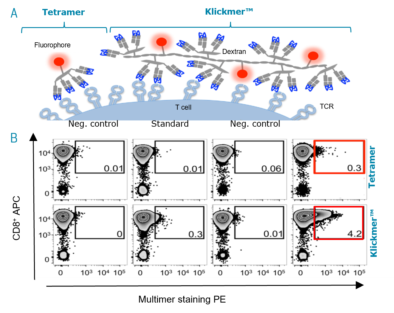
Fig. 1: Detection of preproinsulin (PPI) T cells is further improved using Klickmer® reagents under optimal staining conditions.
A Schematic representation of tetramers and Klickmer® reagents as illustrated by Dolton et al. 2018.
B T cells were also stained with irrelevant peptides as negative control or PPI tetramers (upper panel) and Klickmer® reagents (lower panel) using standard and optimal protocols (PKI + anti-PE Ab). The percentage of cells residing in each gate is shown. The red box indicates tetramer or Klickmer® detected cells.
Conclusions
- The application of Klickmer® reagents extended the TCR affinity threshold amenable to MHC-peptide multimer staining, ensuring clear recovery of the self-reactive, antigen-specific T cells from patient samples by flow cytometry
- Klickmer® further increased the size of the detected population by over 10-fold compared to tetramer staining enabling a higher recovery of the antigen-specific autoimmune T cells.